۱۳۸۹ آبان ۱۱, سه‌شنبه

Sea urchins. کاری از خانم فاطمه عباسی تبار.نام استاد:دکتر مینو محمودی


[edit] Sea urchins
The following description concerns gastrulation in echinoderms, representative of the triploblasts, or animals with three embryonic germ layers. The illustration, however, depicts the gastrulation of a diploblast, animals with two germ layers.
Sea urchins deviate from simple cleavage at the fourth cleavage. The four vegetal blastomeres divide unequally to produce four micromeres at the vegetal pole and four macromeres in the middle of the embryo. The animal cells divide meridionally and produce mesomeres.
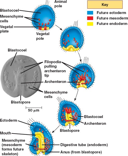
At the beginning of vertebrate gastrulation, the embryo is a hollow ball of cells known as the blastula, with an animal pole and a vegetal pole. The vegetal pole begins to flatten to form the vegetal plate. Some of the cells of the vegetal pole detach and through ingression become primary mesenchyme cells. The mesenchyme cells divide rapidly and migrate along the extracellular matrix (basal lamina) to different parts of the blastocoel. The migration is believed to be dependent upon sulfated proteoglycans on the surface of the cells and molecules on the basal lamina such as fibronectin. The cells move by forming filopodia that identify the specific target location. These filopodia then organize into syncytial cables that deposit the calcium carbonate that makes up the spicules (the skeleton of the pluteus larva).
During the second phase of gastrulation, the vegetal plate invaginates into the interior, replacing the blastocoelic cavity and thereby forming a new cavity, the archenteron (literally: primitive gut), the opening into which is the blastopore. The arechenteron is elongated by three mechanisms.
First, the initial invagination is caused by a differential expansion of the inner layer made of fibropellins and outer layer made of hyalin to cause the layers to bend inward.
Second, the archenteron is formed through convergent extension. Convergent extension results when cells intercalate to narrow the tissue and move it forward.
Third, secondary mesenchyme pull the tip of the archenteron towards the animal pole. Secondary mesenchyme are formed from cells that ingress from, but remain attached to, the roof of the archenteron. These cells extend filopodia that use guidance cues to find the future mouth region. Upon reaching the target site, the cells contract to pull the archenteron to fuse with the ectoderm. Once the archenteron reaches the animal pole, a perforation forms, and the archenteron becomes a digestive tract passing all the way through the embryo.
The three embryonic germ layers have now formed. The endoderm, consisting of the archenteron, will develop into the digestive tract. The ectoderm, consisting of the cells on the outside of the gastrula that played little part in gastrulation, will develop into the skin and the central nervous system. The mesoderm, consisting of the mesenchyme cells that have proliferated in the blastocoel, will become all the other internal organs.

An extracellular matrix molecule that is selectively expressed during development is important for gastrulation in the sea urchin embryo.
Berg LK, Chen SW, Wessel GM.
Department of Molecular and Cell Biology & Biochemistry, Brown University, Providence, RI 02912, USA.
The extracellular matrix is important in the regulation of many cellular events of early development including migration, shape change, proliferation and gene expression. In the sea urchin embryo, disruption of the extracellular matrix results in selective defects in each of these events during gastrulation. Here we describe a new molecule of the extracellular matrix in Lytechinus variegatus, referred to as ECM 18, that has several important features. First, antibody interference of ECM 18 results in a profound but reversible inhibition of primary mesenchyme cell organization and endoderm morphogenesis during gastrulation. Second, during gastrulation, ECM 18 mRNA accumulates to highest levels in the invaginating endoderm and the ECM 18 protein deposited in the basal lamina surrounding the archenteron as well as in other areas of the blastocoel wall. Immunolocalization by fluorescence and electron microscopy demonstrates the selective accumulation of ECM 18 in the extracellular matrix. Third, although the mRNA encoding ECM 18 is present throughout development, the protein accumulates only during gastrulation. ECM 18 protein is not detected in eggs or early embryos and analysis of polysome-associated mRNA suggests that at least part of the translational regulation of ECM 18 is at the level of ECM 18 mRNA-polysome formation. Finally, sequence analysis of ECM 18 shows that the protein contains a repeat sequence with a conserved cysteine motif, suggestive of involvement in protein-protein interactions. Thus, ECM 18 appears to be important in mediating select morphogenetic changes during gastrulation and the pattern of its expression in the embryo is unique among the extracellular matrix molecules known in this embryo.
PMID: 8625821 [PubMed - indexed for MEDLINE]

transcription Factor Activation in the Sea Urchin Embryo
It should be noted that the biochemical nature of the vegetalizing signal(s) has not yet been elucidated. Hörstadius interpreted these experiments in terms of gradients of animalizing and vegetalizing substances. More recently, Davidson (1989) sought to explain these phenomena in terms of transcriptional regulatory proteins (such as those that bind to promoters or enhancers) that are localized throughout the egg and that become activated by inductive interactions between adjacent cells. One of these factors, the one in the micromere precursors of the primary mesenchyme cells, is thought to become active autonomously, early in cleavage. This factor would cause the determination of the micromeres, alter their cell membranes so they could react with the cells above them, and initiate the cascade of determination. By the end of blastulation, there would be five territories that would constitute both the precursor cells for certain regions of the pluteus larva and the regions of specific gene activation. For example, the cytoskeletal actin gene, which is expressed only in the aboral ectoderm cells, cannot be activated anywhere else in the embryo. Similarly, if one injects the gene for the skeletal matrix protein anywhere else in the embryo except the region of skeletogenic mesenchyme, it will not be activated (Hough-Evans et al., 1987; Sucov et al., 1988). Therefore, each territory would constitute a region of differential gene expression that would be reflected in different parts of the organism being formed.
However, if the embryo is perturbed, the neighbors would differ. New fates would ensue as the new vegetal cells activated their skeletogenic mesenchyme determinants and started activation of their neighbors. To see if this was indeed the case, Ransick and Davidson (1993) transplanted micromeres into the animal cap of an 8-cell sea urchin embryo. These skeletogenic mesenchyme precursor cells induced the formation of the archenteron and the expression of the archenteron-specific genes. The fluorescently labeled micromeres did not contribute structurally to the secondary archenteron, but provided a short-range inductive signal that respecified the animal cap cells.
Literature Cited
Davidson, E. H. 1989. Lineage-specific gene expression and the regulative capacities of the sea urchin embryo: a proposed mechanism. Development 105: 421-446.
Hough-Evans, B. R., Franks, R. R., Cameron, R. A., Britten, R. J. and Davidson, E. H. 1987. Correct cell type-specific expression of a fusion gene injected into sea urchin eggs. Dev. Biol. 121: 576-579.
Ransick, A. and Davidson, E. H. 1993. A complete second gut induced by transplanted micromeres in the sea urchin embryo. Science 259: 1134-1138.
Sucov, H. M., Hough-Evans, B. R., Franks, R. R., Britten, R. J. and Davidson, E. H. 1988. A regulatory domain that directs lineage-specific expression of a skeletal matrix protein in the sea urchin embryo. Genes Dev. 2: 1238-1250.
© All the material on this website is protected by copyright. It may not be reproduced in any form without permission from the copyright holder.
HOME :: CHAPTER 8 :: SEA URCHIN CELL SPECIFICATION :: TRANSCRIPTION FACTOR ACTIVATION IN THE SEA URCHIN EMBRYO PREVIOUS :: NEXT

Home Link

ترجمه بخش آخر فصل 9 تکوین.طیبه توسن،ارشد فیزیولوژی.نام استاد:دکتر محمودی


بســــم الـله الـــرحمـن الـــرحیــم

الگو ها(متد ها) شروع بیان ژن هموتیک:

زمینه ی آغازین بیان ژن هموتیک تحت تاثیرژن های gap وparir_rule قرار میگیرد.
به عنوان نمونه:
پروتین های kruppel gap وHunchback ژن های AbdB وabdA را مهار می کند.
این باز دارندگی از شروع بیان ژن های ی که نقش تعیین قسمت های شکمی را دارند در سرو تنه جلوگیری میکند،به عکس oltrabithorax ،توسط سطوح بالایی از Huchbacke فعال کند.ورونویسی Antennapedia توسط Krappleفعال می گردد.
الگوی حفظ بیان ژن هموتیک:
بیان ژن هموتیک فرایندی دینامیکی است. به عنوان نمونه ژن Antp (Antenna pedid) اگرابتدا ،در محدوده ی قطعه ی 4 بیان شود ، خیلی سریع در محدوده ی قطعه ی 5 نیز پدیدار می شود.
مانند germ band گسترش می یابد به طوری که این ژن در لوله عصبی فرضی دور از محدوده ی قطعه ی 12 دیده می شود.
Antp مثل ژن های هموتیک دیگر به صورت منفی تنظیم می شود به بیانی دیگر سرکوب شدن هر یک از ژن های کمپلکس bithorax موجب بیان Antp می شود.
اگر ژن Ultrabithorax را حذف کنیم ،فعالیت Antp گسترش می باشد گر چه در آن قسمت ژن Ubx به صورت نرمال بیان خواهد شد ولی در جایی که بیان Abd شروع می شود متوقف می گردد.(این عمل به دنبال شکل گیری سومین قطعه سینه ای شبیه دومین قطعه ی سینه ای) اگر کمپلکس کامل bithorax را حذف کنیم بیان ژن گسترش می یابد .(چنانکه لاروی باقی نمی ماند اما نمونه کوتیکولی در سرتاسر شکم میباشد که از قطعه ی دوم سینه ای است.)
ژن های Realisutor:
هم اکنون کاووش در مورد ژن های Realisutor است. این ژن ها یاهداف پروتین های همئوتیک اند و عملکرد هر یک به شکل بافت خاص یااندام اصلی است به عنوان مثال برای شکل گیری قطعه دوم سینه ای Antennapodia بیان می شود.
CasaresوMann در سال1998 نشان دادند که پروتین Antennapodia به enhuncer (افزایش دهنده) ژن homotorax وصل می شود و از بیان آن جلوگیری می کند.
Homotoraxبرای ایجاد فاکتور مهم رونویسی تشکیل شاخک ها ضروری است. بنابراین یکی ازعملکردهای Antenna bodia حمایت کردن ژن های ضروری برای رشد شاخک هاست.
پروتین ultrabitorax قادر به ممانعت از بیان زن winglss است این زن در سلول هایی است که به طناب های پروازی تبدیل خواهند شد .
Wingless فقط در سلول های ضمیمه ای شکل دومین و سومین قطعات سینه ای بیان می شود. از کارهای مهم دیگر این ژن پروموتور رشد در این بافتهاست.
در قطعه ی سوم به دلیل وجود wingless Ubdبیان نمیشود که این خود مشخصه ی قطعه ی سوم است.
هدف دیگر پروتین های همئوتیک ژن dital_less کهبای رشد دست و پا ضروری است.
این ژن فقط در بخش سینه ای است و احتمالا در بخش شکمی توسط ترکیبی از پروتین ها ی
Ubx و AbdA ممانعت می شود.
پروتئین های همئودمین:
پروتئین های همئودمین خانواده ای از فاکتورهای رونویسی اند، با دامنه ای از60اسید آمینه مشخص می شونداین پروتئن ها توسط یک توالی با 180جفت باز DNA که همئوبوکس نام دارد بیان می شود.ظاهرا"این پروتئین ها محل اتصال به DNAبوده ودر تعیین نوشت سلول ها مهم وحیاتی اند.
همئودومین در داخل سه مارپیچ قرار دارد.
دو مارپیچ داخل ترکیبی از helix_Turn_helix که ویژگی های فاکتور رونویسی متصل به DNA در شیار بزرگ دوتایی است،قرار دارند.
مارپیچ سوم ،مارپیچ شناسایی است و آن محلی است که اسیدآمینه با پایه های DNA تماس پیدا می کند.
4 باز تکراری TAAt تقریبا در همه ی جایگاه های شناسایی توسط همئودومین حفظ میشود،شاید پروتین های همئودومین این جایگاه ها را می شناسند که می توانند به آن متصل شوند. به نظر می رسد که انتهای '5 در این شناسایی مهم باشد ، جهش در آن اتصال، همئودومین را از بین می برد.
زوجهای اصلی دنبال چهار باز تکراری AttA نیز در تشخیص جایگه های اتصال مهم اند .
مثلا زوج باز بعدی توسط AA مارپیچ شناسایی (مارپیچ سوم) تشخیص داده می شود.مطالعات نشان میدهد که فایده پروتین همئودمین AntennapediaوBicoidT،لیزین وگلوتامین به ترتیب در9جایگاه،مشخص کردن جایگاه های وابسته به شناسایی اند.
لیزین همئو دمین Bicoid،GراازبازCGشناسایی می کند درحالی که گلوتامین همودمین Antenpedia،Aراازجفت بازATتشخیص میدهد.
اگرلیزین درBicoidتوسط گلوتامین جایگزین شود،پروتئین حاصل جایگاه های اتصال Antennapediaراشناسایی خواهد کرد.پروتین همودمین های دیگر نیزالگوهای دیگری رانشان می دهند.یکی از این پروتین ها ی همودمین توالی عادی TAATراشناسایی می کند،درحالی که پروتئین های دیگر ساختارهای مجاور راشناسایی می کنند.
کوفاکتورها برای ژن هایHom –C
ژن های کمپلکس همئوتیک در درزوفیلا سرنوشت قطعه ای را تعیین می کنند اماممکن آنهابرای انجام دادن کارخود به کمک بعضی ژن های دیگر نیزاحتیاج داشته باشند جایگاه های اتصال –DNAهمودمین توسط پروتئین Hom-C شناسایی شده خیلی شبیه اند.دربعضی موارد این اتصالات به دلیل ویژگی های ها یشا ن روی هم قرارمی گیرند
(با هم منطبق اند). درسال 1990PeiferوWieschausکشف کردندکه محصول ژن Exdتوسط چندین پروتئین Hom-C جذب میشوند که ممکن است همانند سازی قطعه ی خاصی کمک کنند.برای مثال Ubxمسئول مشخص کردن قطعه ی همانند شکمی (A1). علاوه بر آن پروتئین ها ی Exdوubx هر دو برای تنظیم ژنdecapentaplegic مورد نیاز می با شند .
پروتئین Extradenticle حاوی یک همئودمین بوده و PBX1 که یک پروتئین انسانیست شباهت زیادی به Extradenticle دارد که ممکن نقش مشابهی به عنوان کوفاکتوربرای ژن همئوتیک انسان بازی کند.
محصول ژن Teashirt نیز به عنوان کوفاکتوردارای اهمیت باشد این فاکتور رونویسی انگشت zincبرای کاهش عملکردپروتئین شانه های جنسی ضروری است.
اگرکمپلکس BithoraxوژنAntennapediaرابردارندهمه ی قطعات پیش سینه ای قدامی می شوند .عامل ریخت زایی قطبیت خلفی-قدامی
قطبیت خلفی-قدامی توسط غلظت فاکتوررونویسی،که Dorsalنام دارد ثابت می ما ند وشبیه Bicoidنیست که غلظت داخل سلول چندهسته ای را برقرار می کند.
تشخیص محور پشتی –شکمی در چندین مرحله صورت می گیرد:
مرحله ی مهم ،تغییرمکان پروتئین Dorsalاز سیتوپلاسم به داخل هسته ی سلول های قدامی که طی تقسیم 14 سلولی انجام می گیرد.آندرسون ونوسلین-ولهارد درسال 1984
11ژن وابسته وموثر را کشف کردند ،که فقدان هریک منجر به کمبود در ساختار قدامی
می شد.مثلا کمبود Cactusباعث شکمی شدن همه سلول ها می شد.
به نظر می رسدکه غلظت های متفاوت پروتئین Dorsalتعین سرنوشت متقاوت درسلول ها داشته باشد.
ترجمه ء پروتئین Dorsal
در واقع این پروتئین dorsa است که بخش پشتی(back ) را از بخش شکمی (belly) معین میکند. رونویسی MNA از ژن dosal مادری در اوویست توسط سلول ها ی تخمدان انجام می شود به علاوه پروتئین تا حدود 90 دقیقه بعد ازر لقاح سنتز نمی شود. وقتی این پروتئین ترجمه می شود آن جزء قسمت پشتی - شکمی در تمام قسمت های جنین یافت می شود.
اما اگر این پروتئین در هر جایی از جنین وجود دارد ،چگونه این پروتئین می تواند به عنوان یک مرفوژن عمل کند؟
جواب شگفت انگیزی در سال 1989 به دست آمد این که، در حالی که پروتئین dorsal می تواند در تمام سیتوپلاسم چند هسته ای پلاستودرم جنین اولیه ء دروزوفیلا یافت شود جزدر هسته هایی که در قسمتهای شکمی جنین واقعند، ترجمه می شوند ،که در آنجا حتما"پروتئین dorsal به ژنهایی متصل می شود که رونویسی را فعال یا متوقف می کنند.
اگر پروتئین dorsal وارد هسته نشود ژنهای مسئول تشخیص انواع سلول های شکمی (twist,snail ) نسخه برداری نمی شوند. وقتی ژنهای مسئول تعیین انواع سلولهای خلفی (decapentaplegic,zerknullt) مهار نشده در نتیجه همه سلولهای جنینی به سلولهای خلفی تبدیل می شوند.
در جهشهایی که همه سلولها خلفی شده اند پروتئین dorsal در هیچ سلولی وجود نداشته برعکس در جهشهایی که در آن همه سلولها فنوتیپ قدامی داشته اند پروتئین dorsal در همه سلولها یافت می شده است.
پیام cascade
پیام از هسته اووسیت به سلولهای فولیکولی:
اگر پروتئین rsal do در تمام قسمتهای جنین یافت شود اما فقط به داخل هسته های سلولهای شکمی وارد گردد و سپس به دیگر سلولها ،باید علامتهای نامتقارنی را به دست آورد. به نظر می رسد که آن سیگنال میانجی باشد اگر چه بر هم کنشی کمپلس بین اووسیت و سلولهای اطراف فولیکولی وجود دارد.
اپیتلیوم اطراف فولیکول اووسیت در ابتدا به صورت متقارن رشد میکند ،اما این تقارن توسط پیام هسته های اووسیت از بین می رود. هسته اووسیت در آغاز در قسمت انتهایی شکمی اووسیت قرار می گیرد دور از سلولهای حامی (nurs). سپس این هسته حرکت می کند و در قسمت پشتی – شکمی قرار می گیرد و سیگنال زیاد سلولهای فولیکولی موجب ستونی تر شدن سلولهای فولیکولی می شود. سیگنال پشتی هسته های اووسیت به نظر تولید ژن gurken را سبب می شود.
پروتئین Gurken به سلول های فولیکولی که به هسته ی اووسیت نزدیکند می رسد.اگر جهش در این ژن ،درداخل اووسیت رخ دهد،باعث قدامی شدن جنین شده وهم سلولهای فولیکولی اطراف آن می شود.اما اگرجهش درسلولهای فولیکولی رخ دهد نه درداخل تخم جنین ایجاد شده طبیعی ونرمال می باشد.
آنالیزمولکولی امروزه نشان داده که همولوگ فاکتوررشداپیدرمی(EGF)مهره داران ژن Gurkenرابه رمزدرمی آورد،درحالی که همولوگ رسپتورEGFمهره داران ژنTorpedo رابیان می کند.کمبودمادرزادی ژنtorpedoموجب قدامی شدن جنین می شود.
Torpedoدرداخل سلولهای فولیکولی تخمدان فعال می شودنه وقتی که جنین شکل گرفت این موضوع با آزمایش زیرثابت شد:
سلول های ژرمینال جنین هایی از تیپ وحشی رابه جنین هایی که مادرشان حامل ژن جهش یافته ی torpedoبودند پیوندزدند،برعکس سلولهای ژرمینال جنین های دارای ژن جهش یافته ی torpedo بودندرا به جنین هایی با تیپ وحشی پیوند زدند.
نتیجه:
تخم های تیپ وحشی داخل فولیکول های مادری حامل ژن جهش یافته رشد کرده وجنین هایی قدامی شده ایجاد شدولی تخم های حاصل از مادرجهش یافته ژنtorpedo توانستندجنین طبیعی ایجاد کنند.
بنا بر این سلولهای فولیکولی به ژن torpedo احتیاج دارند،نه اینکه داخل خود تخم باشد.
سیگنال ازسلولهای فولیکولی به سیتوپلاسم اووسیت:
رسپتور فعال پروتئبن torpedo ازبیان ژ ن pipeجلوگیری کرده درنتیجه پروتئین pipeفقط درسلولهای فولیکولی قدامی ساخته می شود.pipeهم پروتئین Nudalرافعال می کند.
عملکرد سه پروتئازسرین بر روی ژن های easter،snakeو (gd) gastrolation-difectionبه صورت خلاصه:
ابتدا مهارnodal ← فعال شدن gdوشکسته شدن آن ←فعال شدن snake به دنبال آن شکسته شدنr easte ←فعال شدن easter به دنبال آن پروتئین spatzl تقسیم می گردد.اکنون spatzl شکسته شده قادراست به رسپتوری که درغشاءسلول اووسیت وجود دارد باند شود.
ثابت غلظت پروتئین dorsal.
جداسازی پروتئین dorsalوcactus.
ثابت بودن این غلظت چگونه است؟ ظاهر"اپروتئین cactus آن رابلوکه کرده،پروتئینی که dorsal راقادرمی سازدتا به داخل هسته برسد.تا هنگامی که کاکتوس به dorsal اتصال دارد،پروتئین dorsalدر سیتوپلاسم وجود دارد.این سیستم پیچیدهء سیگنالی درجداسازی کاکتوس ازپرو تئین dorsalدرناحیه قدامی تخم سازماندهی می شود: وقتی که spatzlبهtoll متصل می شود←آن را فعال می کند؛.پروتئین کینازpelleرا نیز فعال می کند.فعالیت پروتئین کینازاحتمالا" از طریق یک میانجی پروتئین کاکتوس رافسفریله می کند.فسفریله ی شدن یک بارهءاین پروتئین مقدار آن را کاهش داده←dorsal می تواند واردهسته شود؛که بیشترین آن اغلب درناحیه قدامی ست.
فرایند انتقال پروتئینdorsalبه داخل هسته خیلی شبیه به فرایند انتقال فاکتور رونویسی
B NF-درهسته ی لنفوسیت پستانداران است.
اثرات غلظت پروتئین Dorsal
با نگاهی به نقشه سرنوشت جنین دروزوفیلادربرش عرضی وتقسیم 14 سلول به وضوح دیده می شود که 16 سلول با غلظت با لای پروتئین dorsal مزودرم را ایجاد می کنند؛سلول هایی که درمنطقه بالاتر قرار دارندگلیای تخصصی وسلولهای حد واسط عصبی رو ایجاد می کنند(غلظت dorsalکمتر)و2سلول بعدی اپیدرمیز قدامی وطناب عصبی- قدامی رابه وجود می آورنددر حالی که 9سلول فوقانی تراپیدرمیز خلفی را شکل می دهند؛6سلول هم پوشش آمینوسروزی جنین را ایجاد می کنند.این نقشه بر حسب مقدارغلظت dorsalدرهسته هاست؛مقدار زیاد dorsal موجب القای مزودرمی شدن سلولها می شود درحالی که مقدار کم آن سلولها رابه بافت گلیا یا اکتودرمی شدن سوق می دهد.
اولین اتفاق ریخت زایی درگاسترولاسیون دروزوفیلا پدیده ءتورفتگی سلولها در جنین 16سلولی است که بیشتر ازقسمت قدامی می با شد.ماهیچه های بدن ،گنادها واجسام چرب از سلولهای مزودرمی به وجود می آیند.
پروتئین dorsal مزودرمی شدن این سلول هارا به دوروش معین می –کند.
1) ابتدا فعال کردن ژن های ویژه ای که فنوتیپ مزودرمی ایجاد می کنند.سه ژن هدف عبارت از:
Twist،Snailوrhomboid که فقط درهسته هایی که دارای غلظت بالای پروتئین dorsal اند، رونویسی می شوند.
پروتئین twist ژن های مزودرمی را فعال می کندوsnail از ژن های ویژه ءغیرمزودرمی را که ممکن فعال شوندجلوگیری می کند،rhomboid توسط dorsal فعال اما به وسیله ئ snail ممانعت می شود.درنتیجه rh دراغلب سلولهای قدامی مهار نمی شود.
دو ژنtwistوsnail برای فنوتیپ کامل مزودرمی وگاسترولاسیون طبیعی لازم می باشند.
2) پروتئین dorsal بصورت مستقیم مزودرم را تعین می کند،علاوه برفعال کردن ژن های محرک مزودرمی( snailوtwist ) مستقیما" کار خلفی شدن را انجام می دهند( dppوzen ) ممانعت می کند.درنتیجه این پروتئین می تواند هم به عنوان فعال کننده ی ژن هاعمل کند وهم به عنوان رسپتور ژن های دیگرباشد.
محورها
محورهای پشتی – شکمی وپشتی – جانبی دردروزوفیلا طی یک سیستم هماهنگ شکل می گیرد که می تواند درقسمت های تعین شده ء درون جنین بکار رود.
مطالعات ژنتیکی زیادی روی جنین دروزوفیلا صورت گرفته اماتعداد ژن هایی که تعین محورهای قدامی – خلفی وخلفی – جانبی را عهده دارند کشف نکرده اند.

Enzyme Evolution and Directed Enzyme Evolution


بنام خداوند جان و خرد
كزاين برترانديشه برنگذرد

Enzyme Evolution and Directed Enzyme Evolution


By : MOHAMMAD SADEGH HIRANIAN
Supervised By : Dr MAHMODI

Contents

I. Enzyme Evolution
Introduction
Phylogeny
the Hot Spots and the Cold Spots
Explaining the Hot Spots and the Cold Spots
II. Directed Enzyme Evolution
Introduction
Evolving novel activity
Evolving specific enzymes
Directed evolution usually goes through single amino acid improvements
The challenge of directing evolution
III. Conclusions

Introduction


Importants:

1- reconstructing the historical relationships among various taxa

2- provides an all-important window through which we can glimpse the action of underlying evolutionary mechanisms


continued
Rates of molecular evolution : rates of mutation

rates of spontaneous nucleotide substitution (per site per generation)

low of 2 x 10 -11 in Neurospora crassa
through 5.4 x 10 -10 for Escherichia coli
high of 1.5 x 10 -3 in Qβ.


continued
rates of molecular evolution are greatly modified from the underlying rates of mutation

testify - rates of evolution are presented as substitutions per site per year; rates of mutation are presented as substitutions per site per generation.


Precisely why the rate of molecular evolution should appear so regular on an annual basis, rather than on a per generation basis, remains a mystery.

pseudogenes, evolve slightly faster than introns, 3’- regions of genes and the degenerate sites of coding regions.


The nondegenerate are considered the most constrained because they determine the amino acid sequences of proteins whose functions are directly subject to natural selection.


The frequent replacement of one hydrophobic amino acid by another in the lipid binding domains of mammalian apolipoproteins has been taken as evidence of a lack of structural constraints


The concept of constraint carries force only when supported by evidence garnered independently of evolutionary argument

Constraints are not the only mechanism offered in explanation of rate heterogeneity. Fisher long ago argued that mutations with smaller phenotypic effects are more likely to be selectively advantageous.


rapid evolution seen in degenerate sites within coding regions might be related with smaller phenotypic effects

slow evolution seen at nondegenerate sites is merely a consequence of mutations with larger phenotypic effects being less likely to be advantageous.

Phylogeny


Isocitrate dehydrogenases (IDH) are ubiquitous in nature, catalyzing the oxidation of isocitrate to α -ketoglutarate and CO2 with concomitant reduction of either NAD or NADP.


Many organisms lacking Krebs’ cycle for energy production nevertheless retain its IDH to produce the α-ketoglutarate so essential for glutamate biosynthesis


continued
IDHs belong to an ancient and diverse family of enzymes which include isopropylmalate dehydrogenases, tartrate dehydrogenases, homoisocitrate dehydrogenases

Eubacterial IDHs are approximately 400 amino acid residues long, share at least 40% amino acid sequence identity

Phylogenetic trees have similar topologies.


Most eubacterial IDHs belong to a single monophyletic clade (Figure 1)


the only exceptions being those of Mycobacteria, which more closely resemble the eukaryotic cytosolic IDHs (not shown),

that of Rickettsia prowazekii which more closely resembles the eukaryotic mitochondrial IDHs.


The X-ray structures of Escherichia coli IDH are extraordinarily similar to those of isopropylmalate dehydrogenase, a distantly related member within the protein superfamily.


secondary structure has not evolved within the eubacterial IDH clade, is robustly justified.


the Hot Spots and the Cold Spots


many regions that significant heterogeneity. The most abnormal region is a 10A diameter sphere centered at amino acid residue 153 (Figure 2).

This region evolves at a rate that is 2.8 times slower than the overall average of the molecule. The 59 residue region encompasses much of the active site


Explaining the Hot Spots and the Cold Spots
Directed enzyme evolution Introduction
Unlike rational design, directed evolution does not rely on a detailed understanding of the relationship between enzyme structure and function; rather, it relies on the simple yet powerful Darwinian principles of mutation and selection


Directed evolution is now well established as highly effective for protein engineering and optimization. Directed evolution entails accumulation of beneficial mutations in iterations of mutagenesis and screening or selection


it can be thought of as an uphill climb on a‘fitness landscape’, a multidimensional plot of fitness versus sequence.

Fitness in a directed evolution experiment is defined by the experimenter, who also controls the relationship between fitness and reproduction.


There are an enormous number of ways to mutate any given protein


most mutational paths lead downhill and eventually to unfolded, the challenge lies in identifying an efficient path to the desired function.


Whereas negative epistatic effects are pervasive in natural evolution , such effects have played a major role in facilitating directed evolution.


The vast majority of evolutionary engineering studies over the past ten years involve simple uphill walks, one step at a time.

many ways to create sequence diversity, and this is an important part of the search strategy for molecular optimization.

The goal in choosing a mutagenesis strategy is to minimize the screening requirement and increase the chances of finding beneficial mutations


There is no single ‘best’ mutagenesis method. multiple methods will work (although some are far more efficient than others).


Evolving novel activity

challenge approach to obtaining a new activity was used by Fasan et al. who converted a cytochrome P450 fatty acid hydroxylase into a highly efficient propane hydroxylase, an activity absent in the native enzyme.


Early steps often create ‘generalists’ that are active on a much broader range of substrates. These have been used in a more serendipitous approach to obtaining new activities.


New activities can also arise during ‘neutral drifts’. mutations that do not abolish the native activity are accumulated in multiple rounds of high-error-rate mutagenesis and screening


Evolving specific enzymes


enzyme with high specificity for a new substrate can be generated with a single amino acid substitution.


it may be possible to eliminate variants using negative selection.

Further mutagenesis may also be required to obtain the desired specificity ,with positive selection to improve the desired activity and negative selection to remove the undesired one (s).


Directed evolution usually goes through single amino acid improvements


Analysis of the directed evolution literature shows that a wide range of problems can be solved by uphill walks involving single amino acid changes.


Often, single mutations are responsible for the functional change ,even when multiple mutations are made . Or, when multiple beneficial mutations are found, they all contribute and could have been found separately


Low error-rate random mutagenesis by error-prone PCR is very simple to implement, but only accesses a limited set of (mostly conservative) amino acid changes.


Other mutagenesis methods, including saturation mutagenesis, can effectively generate additional amino acid possibilities in targeted residues .


When the mutation rate is high, beneficial mutations are quickly masked by the much more frequent deleterious ones. Low error rates – 1–2 amino acid substitutions per gene – are therefore preferred if the entire gene is mutated.

Statistical methods to identify beneficial mutations in variants containing multiple mutations have been described and allow simultaneous examination of substitutions at more positions.


The challenge of directing evolution

Even with high-quality mutant libraries and screening, not all bad enzymes can become good ones via a simple uphill walk.


Some problems are more difficult, and coupled mutations might be necessary for the desired functional changes. Beneficial mutations are rare, but combinations of beneficial mutations that only work together are even rarer.


While making targeted libraries may help, it is not clear that we know how to choose the amino acids to target.


It is likely that not all protein folds are ideal for catalyzing desired reactions. Some starting points might require major changes (perhaps even to the protein architecture) in order to improve catalysis and these changes are not accessible by directed evolution.


conclusion

Heterogeneity in rates of molecular evolution

differences in rates of evolution constraints

rates of molecular evolution are greatly modified from the underlying rates of mutation

Directed evolution is now well established as highly effective for protein engineering and optimization

Unlike rational design, directed evolution does not rely on a detailed understanding of the relationship between enzyme structure and function; rather, it relies on the simple yet powerful Darwinian principles of mutation and selection


Supervised By : Dr minoo MAHMODI Basics of Molecular Cloning


کاری از محمد صادق حیرانیان
Defining Cloning
“Cloning” is a loaded term that can be used to mean very different things.
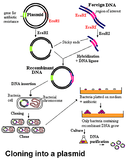
Cutting a piece of DNA from one organism and inserting it into a vector where it can be replicated by a host organism. (Sometimes called subcloning, because only part of the organism’s DNA is being cloned.)
Using nuclear DNA from one organism to create a second organism with the same nuclear DNA

Restriction Enzymes
Restriction Enzymes (also called Restriction Endonucleases) are proteins that cleave DNA molecules at specific sites, producing discrete fragments of DNA.
Restriction Enzymes (RE) were first isolated by Nathans and Smith in 1970.
Why Restriction Enzymes?
Why would bacterial cells contain proteins that cleave DNA at specific sequences?
Generally restriction enzymes are thought to protect bacterial cells from phage (bacterial virus) infection. Bacterial cells that contain restriction enzymes can “cut up” invasive viral DNA without damaging their own DNA.

Joining DNA Fragments
In 1972, Paul Berg and colleagues made the first “artificial” recombinant DNA molecule.
Demonstrated that the DNA of Simian virus 40 could be linearized by EcoR1
Created a circular DIMER of Simian virus DNA by joining two linearized fragments
Also inserted pieces of Lambda phage DNA into linearized Simian 40 virus molecule.

Isolating Genes
Herbert Boyer and Stanley Cohen built on the work of Berg, Nathans and Smith to use restriction enzymes to isolate a single gene, place it into a plasmid vector.
Bacterial cells were then transformed with the recombinant plasmid.

The bacteria host cells replicated the plasmid, producing many copies of the gene, thus amplifying it.

The practical application was that expensive human protein products, like insulin, which were used to treat disease, could eventually be produced from recombinant molecules in the laboratory using bacteria or another host.

Human protein products like insulin could be used in very large quantities from the recombinant molecule. Patients no longer had to use insulin isolated from pigs or cows.

Plasmid Vectors
Plasmids are circular pieces of DNA found naturally in bacteria.
Plasmids can carry antibiotic resistance genes, genes for receptors, toxins or other proteins.
Plasmids replicate separately from the genome of the organism.
Plasmids can be engineered to be useful cloning vectors.

Plasmid Vectors (continued)
Plasmid vectors can be designed with a variety of features:
Antibiotic resistance
Colorimetric “markers”
Strong or weak promoters for driving expression of a protein

Performing the Restriction Digests
You will need to set up a restriction digest of your plasmid vector and your DNA of interest

Restriction enzymes all have specific conditions under which they work best. Some of the conditions that must be considered when performing restriction digest are: temperature, salt concentration, and the purity of the DNA

Purify your DNA Fragments
The insert of interest that you want to clone into your plasmid needs to be separated from the other DNA
You can separate your fragment using Gel Electrophoresis
You can purify the DNA from the gel by cutting the band out of the gel and then using a variety of techniques to separate the DNA from the gel matrix

Ligation
Ligation is the process of joining two pieces of DNA from different sources together through the formation of a covalent bond.
DNA ligase is the enzyme used to catalyze this reaction.
DNA ligation requires ATP.

Transforming Bacteria
After you create your new plasmid construct that contains your insert of interest , you will need to insert it into a bacterial host cell so that it can be replicated.
The process of introducing the foreign DNA into the bacterial cell is called transformation.

Competent Host Cells
Not every bacterial cell is able to take up plasmid DNA.
Bacterial cells that can take up DNA from the environment are said to be competent.
Can treat cells (electrical current/divalent cations) to increase the likelihood that DNA will be taken up
Two methods for transforming: heat shock and electroporation

Selecting for Transformants
The transformed bacteria cells are grown on selective media (containing antibiotic) to select for cells that took up plasmid.
For blue/white selection to determine if the plasmid contains an insert, the transformants are grown on plates .

What did the cells take up?
Plasmid only
Plasmid with insert cloned
Foreign DNA from the environment
Nothing

Expressing your cloned gene
Even if your plasmid contains insert, it may not be able to generate functional protein from your cloned DNA.
The gene may not be intact, or mutations could have been introduced that disrupt it.
The protein encoded by the gene may require post-translational modifications (i.e., glycosylation or cleavage) to function.
Also, some enzymes are a complex of peptides expressed from separate genes.

Expressing your cloned gene
Expression of a cloned gene can be accomplished by:
The E. coli host
Mammalian cells (if the plasmid used is designed for expression in mammalian cells)
Using an in vitro using a cell-free system.

۱۳۸۹ آبان ۹, یکشنبه

گاسترولاسیون توتیای دریایی. دکتر محمودی


An extracellular matrix molecule that is selectively expressed during development is important for gastrulation in the sea urchin embryo.
Berg LK, Chen SW, Wessel GM.
Department of Molecular and Cell Biology & Biochemistry, Brown University, Providence, RI 02912, USA.
The extracellular matrix is important in the regulation of many cellular events of early development including migration, shape change, proliferation and gene expression. In the sea urchin embryo, disruption of the extracellular matrix results in selective defects in each of these events during gastrulation. Here we describe a new molecule of the extracellular matrix in Lytechinus variegatus, referred to as ECM 18, that has several important features. First, antibody interference of ECM 18 results in a profound but reversible inhibition of primary mesenchyme cell organization and endoderm morphogenesis during gastrulation. Second, during gastrulation, ECM 18 mRNA accumulates to highest levels in the invaginating endoderm and the ECM 18 protein deposited in the basal lamina surrounding the archenteron as well as in other areas of the blastocoel wall. Immunolocalization by fluorescence and electron microscopy demonstrates the selective accumulation of ECM 18 in the extracellular matrix. Third, although the mRNA encoding ECM 18 is present throughout development, the protein accumulates only during gastrulation. ECM 18 protein is not detected in eggs or early embryos and analysis of polysome-associated mRNA suggests that at least part of the translational regulation of ECM 18 is at the level of ECM 18 mRNA-polysome formation. Finally, sequence analysis of ECM 18 shows that the protein contains a repeat sequence with a conserved cysteine motif, suggestive of involvement in protein-protein interactions. Thus, ECM 18 appears to be important in mediating select morphogenetic changes during gastrulation and the pattern of its expression in the embryo is unique among the extracellular matrix molecules known in this embryo.
PMID: 8625821 [PubMed - indexed for MEDLINE]

transcription Factor Activation in the Sea Urchin Embryo
It should be noted that the biochemical nature of the vegetalizing signal(s) has not yet been elucidated. Hörstadius interpreted these experiments in terms of gradients of animalizing and vegetalizing substances. More recently, Davidson (1989) sought to explain these phenomena in terms of transcriptional regulatory proteins (such as those that bind to promoters or enhancers) that are localized throughout the egg and that become activated by inductive interactions between adjacent cells. One of these factors, the one in the micromere precursors of the primary mesenchyme cells, is thought to become active autonomously, early in cleavage. This factor would cause the determination of the micromeres, alter their cell membranes so they could react with the cells above them, and initiate the cascade of determination. By the end of blastulation, there would be five territories that would constitute both the precursor cells for certain regions of the pluteus larva and the regions of specific gene activation. For example, the cytoskeletal actin gene, which is expressed only in the aboral ectoderm cells, cannot be activated anywhere else in the embryo. Similarly, if one injects the gene for the skeletal matrix protein anywhere else in the embryo except the region of skeletogenic mesenchyme, it will not be activated (Hough-Evans et al., 1987; Sucov et al., 1988). Therefore, each territory would constitute a region of differential gene expression that would be reflected in different parts of the organism being formed.
However, if the embryo is perturbed, the neighbors would differ. New fates would ensue as the new vegetal cells activated their skeletogenic mesenchyme determinants and started activation of their neighbors. To see if this was indeed the case, Ransick and Davidson (1993) transplanted micromeres into the animal cap of an 8-cell sea urchin embryo. These skeletogenic mesenchyme precursor cells induced the formation of the archenteron and the expression of the archenteron-specific genes. The fluorescently labeled micromeres did not contribute structurally to the secondary archenteron, but provided a short-range inductive signal that respecified the animal cap cells.
Literature Cited
Davidson, E. H. 1989. Lineage-specific gene expression and the regulative capacities of the sea urchin embryo: a proposed mechanism. Development 105: 421-446.
Hough-Evans, B. R., Franks, R. R., Cameron, R. A., Britten, R. J. and Davidson, E. H. 1987. Correct cell type-specific expression of a fusion gene injected into sea urchin eggs. Dev. Biol. 121: 576-579.
Ransick, A. and Davidson, E. H. 1993. A complete second gut induced by transplanted micromeres in the sea urchin embryo. Science 259: 1134-1138.
Sucov, H. M., Hough-Evans, B. R., Franks, R. R., Britten, R. J. and Davidson, E. H. 1988. A regulatory domain that directs lineage-specific expression of a skeletal matrix protein in the sea urchin embryo. Genes Dev. 2: 1238-1250.
© All the material on this website is protected by copyright. It may not be reproduced in any form without permission from the copyright holder.
HOME :: CHAPTER 8 :: SEA URCHIN CELL SPECIFICATION :: TRANSCRIPTION FACTOR ACTIVATION IN THE SEA URCHIN EMBRYO PREVIOUS :: NEXT

Home Link

گاسترو لاسون درتوتیای دریایی

توضیحات زیر مربوط به گاسترولاسیون در خارپوستان است .(جاندارانی با 3 لایه ی جنینی) هرچند توضیحات مربوط به تصویر گاسترلاسیون جانورانی است با دو لایه ی جنینی.


توتياي دریایی تسهیم کامل داردتسهیم اول ودوم هر دو نصف النهاری و عمود بر هم می باشد. تسهیم سوم مداری وعمود بردو صفحه ی تقسیم اول ودوم انجام شده قطب جانوری وگیاهی را از هم جدا می کند تسهیم چهارم کاملا متفاوت از سه تسهیم اول می باشد درتوتیای دریایی از کلیواژ ساده به طرف شکافتگی چهارم (پیشرفته تر ) هدایت شده ، چهار بلاستومر قطب نباتی به طور نامساوی تقسیم شده ، 4 میکرومر را در نزدیکی قطب نباتی و 4 ماکرومر را در بخش میانی ایجاد کرده سلول های قطب حیوانی نیز تقسیم نصف النهاری انجام داده و مزومرها را تولید می کند .
مرحله ی بلاستولای تکوین در توتیای در یایی از مرحله 128 سلولی شروع می شود.در این زمان سلول ها کرهی تو خالی را بوجود می آورند که حفره مرکزی بلاستوسل را در برمی گیرد.که حاوی مایع پروتیینی می باشد.بلاستومرها اطراف این حفره با اتصالاتی محکم صفحه اپی تلیالی پیوسته ای را ایجاد می کند.همچنین هر یک از سلولها (بلاستومرها)دربخشی از غشای سلولها که دور از حفره مرکزی بلاستوسل قرار دارد مژه دار می شود و بلاستولای مژه دار در پوشش لقاحی خود شروع به چرخیدن کرده ومتعاقب آن تمایز می یابند.
جنین در قطب نباتی شروع به پهن شدن کرده و به شکل پلاک نباتی در می آید ،برخی از سلول ها از قطب نباتی مهاجرت کرده وپس ازعبورتبدیل به سلولهای مزانشیمی اولیه می شوند ،سلول های مزانشیمی اولیه تقسیم شده و در امتداد ماتریکس خارج سلولی به طرف بخش های مختلف بلاستوسل می روند .
این مهاجرت بنابر اعتقادی بستگی دارد به پروتئوگلیکان سطح سلول ها و ملکول هایی بر روی غشای پایه که فیبرونکتین نامیده می شوند . حرکت سلول ها توسط فیلوپودیاها هدایت می شود به عبارتی این فیلوپودیاها محل سلول هدف را می شناسند.
ساختار (گودالی)به نام آرکنترون را ایجاد می کند که در دهانه ی آن بلاستوپور نامیده می شود .
توسعه آرکنترون توسط 3 مکانیسم ادامه می یابد:
1)درون خزیدگی اولیه ایجاد می شود . با انبساط پی در پی لایه ی درونی ساخته شده از فیبروپیلن و لایه ی خارجی ساخته شده از هیالین.
2)آرکنترون شکل می گیرد از توسعه ی همگرا (توسعه ای که به همه ی قسمت ها برسد ) . توسعه ی همگرا زمانی اتفاق می افتد که سلول ها در میان بافت باریکی جای گرفته و این ساختار به جلو حرکت می کند .
3)مزانشیم ثانویه کشیده می شود به نوک آرکنترون . مزانشیم ثانویه شکل می گیرد از سلول هایی که وارد می شوند و متصل به سقف آرکنترون باقی می مانند .

فیلوپودیا راهنمای هدایت است . که بعداً این ساختار در ناحیه دهان یافت می شود به محض رسیدن به هدف فیلوپودیا با اکتودرم تماس می یابد و سلول مزانشیم به قطب حیوانی می رسد و در نهایت یک حفره ی گوارشی ایجاد می شود که از همه ی راههای میان
جنین می گذرد .
4)لایه های 3 گانه ی جنینی شکل می گیرد . اندودرم به قطعه ی گوارشی ، اکتودرم شامل سلول های خارج گاسترولاست که در گاسترولاسیون نقش کمی ایفا میکند واین اکتودرم به پوست و سیستم عصبی مرکزی توسعه می یابد، مزودرم شامل سلول های مزانشیمی است که در بلاستوسل توسعه می یابد و تبدیل به اندام داخلی جنین می شود


مهاجرت انفرادی مزانشیم اولیه
عملکرد سلولهای مزانشیمی اولیه .مدت زمان کوتاهی پس از خروج بلاستومرها از پوشش لقاحی گروهی از سلولهای مشتق شده از میکرومرها نوعی تغیر شکل از حالت اپی تلیا ل به مزانشیمی پیدا می کنند.این سلولها اسکلت سلولی خود را تعغیر داده و بطری شکل می شوند همچنین اتصال خودرابا سلولهای جانبی از دست داده و با جدا شدن از لایه راسی وارد بلاستوسل می شوند.(شکل15-8ساعت 10-9)
این سلولهای مشتق شده از میکرومرها مزانشیم اولیه نامیده می شوند.از انجایی که این سلولها اسکلت لا روی راتشکیل میدهند و مزانشیم اسکلت زا نیز گفته می شود این سلولهای مزانشیمی قادرند زائدهایی بلند و نازک و منقبض شونده (به نام فیلو پودیا را به وجود اورند.

.در ابتدا به نظر می رسد که این سلولها به صورت تصادفی در امتداد سطح درونی بلا ستوسل حرکت می کنند.برای انجام حرکت زوائد فیلو پود یایی این سلو لها به طور متناوب اتصالاتی را با دیواره ی بلاستوسل برقرار کرده وسپس جدا می شوند نهایتا این سلولها در ناحیه شکمی – جانبی اینده ی بلاستوسل مستقر میگردند.در این مکان به طناب سین سیشیومی که در تشکیل محورکربنات کلسیمی اسپیکول های اسکلتی لارو نقش دارد ملحق می شوند (شکل d-e14-8)


شكل 8-15


اهمیت لایه ی ماده ی زمینه ای برون سلولی درون بلا ستوسل.
مهاجرت نسل میکرمری به درون بلا ستوسل در نتیجه ی از دست رفتن تمایل این سلولهای مزانشیمی اولیه برای اتصال به سلولهای مجاور و غشای هیالینی ودر عوض تمایل بالای انها برای اتصال یافتن به گروهی از پروتئینها که دربلاستوسل قرار گرفته اند رخ می دهد این نوع مدل مهاجرت مزانشیمی برای اولین بارتوسطgustafsonوwolpertدر 1967 ارائه شد ودر سال 1985زمانی که rachelfinkوdavidmcclay میزان قدرت اتصال بلا ستومرهای توتیایی دریایی به لایه ی هیا لینی غشای پایه ی بلاستوسل وسایر بلاستومرها رااندازه گیری کردند تایید گردید.

تمامی سلولهای بلاستولا در ابتدا از سطح بیرونی خود به لایه ی هیالینی و از سطح درونی خود به غشای پایه ی ترشح شده توسط خودشان متصل هستند .هر سلول از سطوح جانبی نیز به سلولهای همسایه اتصال دارد.finkوmcclayدریافتند که سلولهای اکتودرمی و اندودرمی اینده ی جنین (به ترتیب اعقاب مزومرها وماکرومرها)محکم به یکدیگر و به لایه ی هیالینی متصل می شوند اما اتصال این سلولها به غشای پایه سست است میکرومها نیز در ابتدا الگوی اتصالی مشابهی را دارند اما الگوی انها حین گاسترو لاسیون تغییر می کند در حالی که سلولهای دیگر اتصال محکم خود را با لایه ی هیالینی و سلولهای مجاور حفظ می کنند پیش سازهای مزانشیمی اولیه تمایل خود را نسبت به این ساختارها از دست می دهند (این تمایل به حدود 2درصد تمایل اولیه می رسد)اما تمایل انها نسبت به ترکیبات موجود درغشا ی پایه و ماده ی زمینه ای برون سلولی (نظیر فیبرونکتین)صد برابر بیشتر می شود


تمامی سلولهای بلاستولا در ابتدا از سطح بیرونی خود به لایه ی هیالینی و از سطح درونی خود به غشای پایه ی ترشح شده توسط خودشان متصل هستند .هر سلول از سطوح جانبی نیز به سلولهای همسایه اتصال دارد.finkوmcclayدریافتند که سلولهای اکتودرمی و اندودرمی اینده ی جنین (به ترتیب اعقاب مزومرها وماکرومرها)محکم به یکدیگر و به لایه ی هیالینی متصل می شوند اما اتصال این سلولها به غشای پایه سست است میکرومها نیز در ابتدا الگوی اتصالی مشابهی را دارند اما الگوی انها حین گاسترو لاسیون تغییر می کند در حالی که سلولهای دیگر اتصال محکم خود را با لایه ی هیالینی و سلولهای مجاور حفظ می کنند پیش سازهای مزانشیمی اولیه تمایل خود را نسبت به این ساختارها از دست می دهند (این تمایل به حدود 2درصد تمایل اولیه می رسد)اما تمایل انها نسبت به ترکیبات موجود درغشا ی پایه و ماده ی زمینه ای برون سلولی (نظیر فیبرونکتین)صد برابر بیشتر می شود

این تغییر در تمایلات سلولی موجب ازادسازی اتصالات سلولی میکرومرها از لایه ی هیالینی خارجی و سلولهای مجاور شده و باعث می شود که از طریق غشای پایه به داخل کشیده شده وبه درون بلاستوسل مهاجرت کنند (شکل a 16-8) این تغییر در تمایلات به دلیل تغییر در مولکولهای سطح سلولی است که در این زمان ایجاد می شودهمچنین پرو تئین هایی نظیر فیبرونکتین اینتگرین لامینین1وکادهرین ها نیز در این مهاجرت سلولی شرکت دارند .
همان گونه که درشکل cوb16-8نشان داده شده غلظت بالایی از مواد برون سلولی در اطراف سلولهای مزانشیمی اولیه ی در حال مهاجرت دیده می شود.در داخل بلاستوسل به نظر می رسید که سلولهای مزانشیمی اولیه در امتداد ماده زمینه ی برون سلولی دیواره بلاستوسل مهاجرت کرده و فیلوپودیای خود رابه سمت جلو گسترده می کنندچندین نوع پروتئین(نظیر فیبرونکتین و نوع خاصی گلیکوپروتئین سولفاته)
در شروع و حفظ این مهاجرت لازم و ضروری هستنداما این علایم جهت یابی کافی نیستند چرا که سلولهای در مهاجرت((میدانند))که چه موقع حرکت خود را متوقف کنند و اسپیکولها را نزدیک استوای بلاستوسل تشكيل دهند.
سلولهای مزانشیم اولیه به صورت یک حلقه در موقعیتی خاص و در امتداد محور جانوری-نباتی سازمان دهی میشوند-بسیاری از سلولهای مزانشیم اولیه در دو ناحیه نزدیک بخش شکمی اینده ی لارو تجمع
یافته با یکدیگر ترکیب شده و شروع به تشکیل اسپیکول می کنند(شکل17-8)


شكل 8-17

اگر میکرومر نشان دار شده ای از یک جنین به بلاستوسل جنین دیگری در مرحله ی گاسترولاسیون تزریق شود به محل صحیح مهاجرت کرده ودر تشکیل اسپیکول های جنینی شرکت می کندتصور می شود که اطلاعات مکانی مورد نیاز توسط سلولهای اکتودرمی اینده وغشا ی پایه ی انه فراهم می شود (شکلa18-8}
تنها سلولهای مزانشیمی اولیه (ونه انواع دیگر سلولها)قادرند به این علایم الگوبندی پاسخ دهند


شكل 8-18


.

millerوهمکارانش در 1995از وجود فیلوپودیا کاملا واضح (با قطر3/0ميكرو متر)در سلولهای مزانشیمی تشکیل دهنده ی اسکلت سلولی خبر دادند
به نظر می رسد که این فیلو پودیا در حرکت نقش فعالی نداشته بلکه به جستجوی دیواره ی بلاستوسل می پردازد وشاید در پاسخگویی به علایم الگو بندی پشتی – شکمی و جانوری نباتی صادر شده از اکتو درم نقش داشته باشد.

مرحله ی اول درون روی آرکنترون
هم زمان که سلول های مزانشیمی اولیه ی قطب نباتی جنین کروی را ترک می کنند ، تغییرات مهمی در سلول های باقی مانده ایجاد می شود.سلول های باقی مانده ضخیم و پهن شده و صفحه ی نباتی را تشکيل دهند.در نتیجه شکل جنین تغییر می کند شكل8 -51)ساعت 9سلول های صفحه ی نباتی به یکدیگر و به لایه ی هیالینی سلول تخم متصل باقی مانده و به گونه ایی حرکت میکنند تا فضاهای خالی به وجود آمده ی حاصل از مهاجرت مزانشیم اولیه را پر کنند. به علاوه صفحه نباتی به درون خم شده وپس از درون روی حدود 4/1الی 2/1به طور ناگهانی متوقف می شوند دهانه این ناحیه درون رفته (آرکنترون ) بلاستو پور نامیده میشود.
بررسیها نشان داده که لایه هیالینی موجوددر صفحه نباتی به دلیل تغییر در ترکیباتش به سمت درون خمیده میشود.لایه هیالینی درواقع از دو تیغه تشکیل شده است یک تیغه بیرونی متشکل از پروتیین هیالین و یک پروتیین درونی متشکل از پروتیین های فیبروپیلین .فیبروپیلین ها در گرانول های ترشحی تخمک ذخیره شده وپس از اگزوسیتوز گرانول های قشری ترشح می شوند و منجر به آزاد سازی پروتیین هیالین می گردند.درمرحله بلاستولا فیبروپلین ها شبکه ای تور ماننددر اطراف جنین ایجاد میکند .در حین درون روی سلول های صفحه نباتی شرو به ترشح گلیکو پروتیین کندرویتین سولفات به درون تیغه درونی لایه هیالینی میکند در نتیجهناحیه نباتی لایه هیالینی خمیده میشود اندکی بعد یک نیروی ثانویه که از حرکت سلولهای اپی تلیالی مجاور صفحه نباتی حاصل میشود با کشیدن لایه خمیده به درون موجب تسهیل در فرآیند درون روی می گردد.


سلول های مزانشیم ثانویه اولین گروه از سلول هایی هستند که درون روی نموده ، به درون بلاستوسل پیش روی می کنند و راس آرکنترون را تشکیل می دهند . آنها نهایتاً سلول های رنگدانه دار ، عضلات اطراف لوله ی گوارشی ، و کیسه های سلومی را تشکیل
می دهند سلول های اندودرمی مجاور مزانشیم مشتق شده از میکرومر ،لوله ی گوارشی قدامی را به وجود می آورند که فاصله ی بیشتری را در بلاستوسل مهاجرت می کند . لایه ی بعد سلول های اندودرمی لوله ی گوارشی میانی را به وجود می آورد . ردیف محیطی آخر نیز درون روی نموده ، لوله ی گوارش خلفی و مخرج را تشکیل می دهد


مراحل دوم و سوم درون روی آرکنترون

دنبال درون روی اول ، فاز دوم شکل گیری آرکنترون شروع می شود . طی این مرحله آرکنترون گسترده شده به گونه ایی که حتی ممکن است طول آن سه برابر شود . در طی این گسترده
شدن لوله ی گوارش کوتاه و عریض به لوله ای باریک و طویل تغییر شکل می دهد . برای این طریق مهاجرت بر روی یکدیگر و پهن شدن باز آرایی می شوند .
پدیده ایی که طی آن سلول ها در هم فرو می روند و موجب نازک شدن بافت شده و هم زمان یه سمت جلو حرکت می کنند بسط همگرا نامیده میشود .

در تمام گو نه های توتیای در یایی مرحله سوم طویل شدن آرکنترون نیز دیده می شود این مرحله انتهایی در اثر نیروی کششی که توسط سلولهای مزانشیم ثانویه ایجاد میشود آغاز میگردد .این سلولها راس آرکنترون را ساخته ودر همان جا مستقر میشوند سلولهای مذکوردر مایع بلاستوسل زواید فیلوپودیایی ایجاد می کنند که به طرف سطح درونی دیواره بلاستوسل کشیده میشود.فیلوپودیاهادرناحیه اتصالی بین بلاستومرهابه دیواره متصل شده سپس با کوتاه شدن خود آرکنترون را به بالا میکشند(شکل های 8-15و 8-21 ساعت 12و13)
. اگرتعدادی از سلول های مزانشیم ثانویه حذف شوند طویل شدن ادمه می یابد، اگرچه سرعت آن کند می شود . در این گونه سلول های مزانشیم ثانویه نقشی اساسی در کشیده شدن آرکنترون به سمت دیوار بلاستوسل حین مرحله ی انتهایی درون روی بازی می کنند. اما آیا فیلوپودیای مزانشیم ثانویه می تواند به هر بخشی از دیواره ی بلاستوسل متصل شود، یا این که هدف خاصی در نیم کره جانوری باید وجود داشته باشد تا اتصال صورت گیرد؟ آیا بخشی از دیواره ی بلاستوسل وجود دارد که متعهد شده تا بخش شکمی لارورابه وجود آورد؟ مطالعات صورت گرفته توسط Hardin وMcCIay در1990 نشان می دهد که ناحیه ی هدف ویژه ای برای فیلوپود یا وجود دارد که از سایر نواحی قطب جانوری متفاوت است فیلوپود یاگسترش می یابد و به صورت تصادفی به دیواره ی بلاستوسل متصل شده وسپس آزاد می گردد. با این وجودزمانی که فیلوپود یا با ناحیه ی خاصی از دیواره برخورد می کند اتصال خود را حفظ کرده ، منقبض شده وآرکنترون را به سمت خود می کشد. زمانی که Hardin وMcCIay سمت دیگر دیواره ی بلاستوسل رادرمعرض فیلوپود یاها قراردادند، فیلوپود یا به گسترش خود ادامه داده وبعد از برخورد با این دیواره ی دست کاری شده آن را رها نمودند. تنها زمانی که فیلوپود یا بافت هدف مورد نظر را پیدا کرد، حرکات خود را متوقف نمود

. اگرگاسترولا به اندازه ای طویل می شد که فیلوپود یا هرگزنمی توانست به ناحیه ی هدف برسد، سلول های مزانشیم ثانویه به جستجوی خود ادمه می دادند وسرانجام آرکنترون را رها کرده وبه صورت آزاد، هدف خود را جستجو می کردند. بنابراین،آنچه واضح است این است که در ناحیه ای که قرار است بخش شکمی لارورا به وجود آورد وهمچنین ناحیه ای از آکنترون که دهان آینده ی جنین را به وجودخواهد آورد،
بخش هدفی وجود دارد که توسط سلول های مزانشیم ثانویه شناسایی می شود . هم زمان با برخورد راُس آرکنترون با ناحیه ی هدف دیواره ی بلا ستوسل سلول های مزانشیم ثانویه روی بلاستوسل پخش می شوند، سپس تکثیر شده واندام های مزودرمی را به وجود می آورند (شکل 15-8 ساعت 5/13). جایی که آرکنترون با دیواربرخورد می کند، نهایتاً به دهان موجود تبدیل خواهد شد
سلول ها مزانشیمی اسکلت زاوارد rudiment شده تا صفحات اسکلتی اولیه سند شوند سمت چپ پلوتئوس سطح دهانی آینده ی جنین توتیای دریایی بالا را به وجود می آورد، حین دگردیسی، imaginal rudime لاروجدا شده سپس لاروتخریب می شود،درحالی که naginal rudiment ( که حالا جوانه نامیده می شود) مجد داً لوله ی گوارش خود ایجاد کرده ودر کف اقیانوس قرار می گیرد. همچنان به مواد غذایی که لاروها شده دریافت کرده وابسته است. الگوی گاسترولاسیون خاربوستان نمونه ی تکاملی تکوین پس دهانیازبه
حساب می آید.درپس دهانیان (خارپوستان ، تونیکات ها سفالوکوردات ها و مهره داران) منفذ اولیه تبدیل به مخرج می شود، درحالیکه منفذ دوم دهان را تشکیل می دهد (دوتروستوم به معنی دهان ثانویه). به علاوه درتوتیای
دریایی پدپده ی گسترش همگرا واستفاده ازبیان ژن Nodal درتثبیت محورها دیده می شود

بلوغ اووسیت و رشد جنین مرغ .استاد : سرکار خانم دکترمحمودی .تحقیق توسط : نرگس آرین جاه


آنچه هستی ، هدیه خداوند است به تو
و
آنچه میشوی ،هدیه توست به خداوند
پس
بی نظیر باش ، زیرا بی نظیر آفریده شده ای
دانشگاه آزاد اسلامی
گروه زیست شناسی
استاد : سرکار خانم دکترمحمودی
تحقیق توسط : نرگس آرین جاه
زیست تکوینی جانوری

The initial stages of development depend on m-rna and proteins accumulated in the oocyte , and during these stages , certain genes are essential for fertilization , first cleavage and embryonic genome activation .
The aim of this study was first to search for avian oocyt – specific genes
The activation of molecular pathways underlying oocyte to embryo transition (oet) depends exclusively on maternal Rnas and proteins accumulated during growth of the oocyte .
Indeed,by the two cell stage , the major pathways regulated by maternal mrna are targeted protein degradation, translational control and chromatin remodelling .
The recruitment of maternal mrna for translation has long been recognized as a widespread mechanism to generate newly synthesized proteins in maturing oocytes and fertilized eggs .
Conversely,rna that is not larger needed in the early embryo.
moreover, careful regulation of proteolysis during the same period is likely to be important in oocytes, which are prodominantly transcriptionally inactive and must often wait for long periods before fertilization in different species.
Maternal transcripts that are present in the early pre-implantation embryo can be subdivided into two classes according to whether they are synthesized soon after embryonic genome activation or not.
The firstis common to the oocyte and early embryo and is replenished after activation of the zygotic genome. The second consists of oocyte-specific mrna that is not subeqently transcribed from zygotic genes in the embryo this class of mrna may be detrimental to early post-fertilization development.
Birds represent a good model to observe progressive accumulation of mrna in the oocyte befor ovulation. The embryonic genome of a model bird(the chicken), is activated when the embryo contains 30000-50000 cells 24h after fertilization.
Protein and mrna, accumulated as the chicken oocyte matures,are essential not only for fertilization and first cleavage but also for supporting a high number of emberyonic cell divisions before genome activation.
The avian oocyte consists of a large amount af yolk and a structure called the germinal disc(gd). The gd is a white plaque of about 3-4mm diameter an the top of the oocyte. It contaions the nucleus and 99% of oocyte organelles althoughit occupies less than 1% of the cell volume.

تعيين جنسيت در پرندگان.ارائه:شهین شبرندی . استاد:دکتر محمودی


تعيين جنسيت در پرندگان
جنس تمام پرندگان از جمله شتر مرغ( ازگونه سينه پهن) به وسيله کرمزومهاي جنسي معين مي گردد . پرندگان نر داراي دو کرمزوم جنسي خاص خود هستند که Z ناميده مي شود . ( ZZ ) ماده ها داراي يک کرمزوم Z و يک کرمزوم W مي باشند . (ZW ) هنگامي که پرنده نر اسپرم توليد مي کند يک کرمزوم Z در اسپرم قرار مي گيرد . اما نقش ماده در هنگام توليد تخم متفاوت است . اگر کرومزوم W در تخم قرار گيرد جنيني که به وجود مي آيد ( ZW ) ماده خواهد بود . در صورتي که تخم داراي کرمزوم Z باشد ، جنين ( ZZ) نر خواهد بود . در مورد شتر مرغها و تقريباً همه پرندگان بر عکس انسان ، کرمزومهاي ماده تعين کننده جنس فرزندان مي باشد .
ژن (DMRT1 ) در پرنده ی سینه پهن ، نقش آن در تعیین جنسیت در پرندگان
خلاصه : بر خلاف پستانداران، پرندگان نر دارای کروموزوم zz و جنس مادهZW می باشند در بیشتر پرندگان کروموزوم z بزرگ و غنی است در حالیکه کروموزوم w کوچک و هتروکروماتینی است . هر ژن متمایز در سینه پهن zو w هستند بنابراین داوطلب قوی برای تعیین جنسیت هستند ، بخش داوطلب تعیین جنسیت در پرندگان ژن1 DMRTدر شتر مرغ استرالیایی (یکی از گونه های پرنده ی سینه پهن) می باشدکه درطول مراحل تمایز غده جنسی در جوجه بیان شده ، برطبق نقشه های ژني این ژن 1DMRT در ناحیه ديستال بازوی کوتاه Z وجود دارد و درروی کروموزوم W دیده نمی شود0 بطور غیره منتظره ،ترادف و توالی ژن DMRT1 شترمرغ استرالیایی 88 درصد همسانی دارد با توا لی DMRT1 موجود در جوجه و 65/0 هم همسانی با توالی ژن 1 DMRT موجود درانسان دارد ، یک ناحیهbp 270 جفت باز بیان نشده در اینترون3 ژن DMRT1شترمرغ استرالیایی که 90/ 0که همسان باتوالی وترتیب در اینترون مربوط به ژن DMRT1در انسان ،این حفاظت فوق العاده بالادرحدود300 میلیون سال از تکامل ، حاکی از یک تابع مهم است در کنترل بیان ژن DMRT1 و تعیین جنسیت درمهره داران 0
ژن DMRTبرای اولین بار در تعیین جنسیت مهره داران دخیل بود که این ژن توسط شناسایی در کروموزوم شماره9 انسان ، این ژن کد می کند پیام را که حاوی پروتئین می باشد که درتعیین جنسیت دخیل می باشد0
درموش خانگی ژن DMRT1 بطور وسیع درستیغ تناسلی هردوجنسxxوxy بیان می شود که درجنس نر درطی
گونادوژنردرلوله های منی ساز بیضه و سلولهای سرتولي دیده شده است وهمچنین درسلولهای بنیادین این ژن وجود دارد 0
پرندگان سیستم تعیین کننده ی کروموزوم جنسی دارند که درپرندگان نر zz ودرپرندگان ماده zw است کروموزوم شماره 9انسان باکروموزوم z درجوجه همسان است ،که ژن DMRT درانسان برروی كروموزوم شماره 9انسان می باشد ودرپرندگان برروی كروموزمZ می باشد ودركروموزم w درپرندگان وجودندارد0
واکنش RT-PCR که شامل CDNAوپرایمرهای DMEX3F1، DMTNE1، DMTNF2،DMTNR1، DMEX4R1 و همچنین dctp،dATP،dTTP و DNAپلیمراز دراین آزمایش (RT-PCR)PCRژن DMRT1 بیان شده در مرزتناسلی متمایز غده جنسی رانشان داده است0 تفاوت در بیان ژن DMRT1 باعث بوجود آمدن دوجنس نر و ماده می شود شکل(3) موقعیت پرایمرهای RT- PCR دراگزون DMRT1 و قطعه اینترون حفاظت شده،اگزون 4 رانشان می دهد.اینترون 3 حفظ شده ناحیه اي مسئول برای تنظیم فعالیت ژن DMRT1 می باشد و یک نقش مهم آن درتنظیم دوزژن تولیدی درپرندگان و بنابراین درتنظیم معیین جنسیت می باشد سینه پهن بزرگ وپرنده ای بدون پرواز است که در میان گونه های آن می توان شترمرغ آفریقایی و شترمرغ استرالیایی رانام برد آنها دارای کرو موزوم z که ازاندازه یکسان با کروموزوم موجه است وکارپوتیپ كروموزوم نشان می دهد که دو کروموزوم جنسی ازنظرتمام طول مشابه اند،برخلاف بخش هتروکروماتیني کروموزوم کوچکw درجوجه ،کروموزوم w شترمرغ استرالیایی بزرگ و جفت با z در بالاترین طول،کارپوتیپ کروموزومی z- DNA جوجه موید آن است کهZ شتر مرغ استرالیایی وWهمسان هستند اما ناحیه ای ویژه ی کوچک درWنزدیک سانترومراست که ماپیش بینی کردیم که این ناحیه ویژه درماده نشان دهنده ی تمایز ابتدايي zw رادرپرندگان مي باشد
بحث:
ما نتیجه می گیریم که ژن DMRT1 روی کروموزوم Z شترمرغ استرالیایی وجود دارد وکپی درکروموزوم W وجود ندارد و همچنین کشف کردیم توالی و ترادف حفاظت شده دردرون اینترون 3 ژن DMRT1 که این توالی اینترون 3 مسئول تنظیم بیان ژن DMRT1 می باشد 0
همسانی کروموزوم 9 انسان باکروموزوم Z جوجه موید این نکته است که ژن DMRT1 یک ژن حیاتی برای تعیین جنسیت درپرندگان می باشد و بیان DMRT1 درطي گنادوژنزدرجنین جوجه با این فرضیه مطاقبت دارد برطبق مشاهده ی نقشه های ژن DMRT1 درکروموزوم Z وعدم وجودآن برروی کروموزوم w درانسان دو نسخه ژن DMRT وجود دارد که برای تعیین بیضه ها لازم می باشند درکروموزوم ZZ پرنده نرژن DMRT1 فقط ازجمله هزاران ژن هایی است که روی کروموزوم Z وجود دارد که نظیر و همانند آن روی کروموزوم هتروکروماتینی کوچک w وجودندارد ودرصورت نبودن جهش وانتقال ژني ، ژن DMRT1درتعیین جنسیت پرندگان نقش دارد .
تعيين جنسيت ژنتيكي در شترمرغ
تعين جنسيت از طريق دي ان آ در شتر مرغها فقط نيازمند يک قطره خون است که از آن يک نمونه DNA استخراج مي شود . پس از طي چندين فرآيند شيميايي ، يک بر چسب راديو اکتيو ( به نام پروب 10 ) به زنجيره DNA چسبانده مي شود . که به دنبال بندهاي DNA در کرمزومهاي جنسي گشته و به آن مي چسبد . پروب نوع کرومزوم هاي جنسي ZZ يا ZW ( يعني نر يا ماده بودن پرنده را تعيين مي کند.يکي از بهترين جنبه هاي تعيين جنسيت از طريق DNA دقت بالاي آن است ( 99 درصد .)
تعيين جنسيت قناري با استفاده از روش PCR
چکيده: تعيين جنسيت اغلب پرندگان تا زمان بلوغ و حتي گاهي بعد از بلوغ امري نسبتا مشکل است. اما جنسيت پرندگان و ساير جانوران از بدو تشکيل نطفه، به لحاظ ژنتيکي معين است. امروزه با پيشرفت روش هاي مولکولي، تعيين جنسيت جانداران در هر مرحله از زندگي ميسر مي باشد. در تحقيق حاظر تعيين جنسيت در قناري با استفاده از واکنش PCR و براساس دو ژن CHD-W و CHD-Z شرح داده شده است. در اين تحقيق جمعا از 29 قطعه قناري استفاده شد. استخراج DNA از انتهاي کالاموس پر قناري هاي زنده صورت پذيرفت و با استفاده از پرايمرهاي اختصاصي طراحي شده در اين طرح، واکنش PCR تنظيم گرديد. نتايج نشان دهنده تکثير دو قطعه DNA به طول هاي 345 و 306 جفت باز براي جنس ماده و فقط يک باند 306 جفت باز براي جنس نر بود. اين نتايج براي كليه نمونه هاي پر مشاهده شد. از آنجا که طراحي اين سيستم تعيين جنسيت براي اولين بار در قناري مورد استفاده قرار مي گيرد و نتايج مولکولي با ويژگي هاي مورفولوژيک دو جنس نر و ماده کاملا مطابقت دارد، لذا تعيين جنسيت جوجه هاي قناري با انجام واكنش PCR روي نمونه هاي پر، آسان، سريع، بي خطر و ارزان مي باشد.


DMRT1 in a ratite bird: evidence for a role in sex determination and discovery of a putative regulatory element


Abstract.
Unlike mammals, birds have a ZZ male/ZW female sex-determining system. In most birds, the Z is large and gene rich, whereas the W is small and heterochromatic, but the ancient group of ratite birds are characterized by sex chromo­somes that are virtually homomorphic. Any gene differentially present on the ratite Z and W is therefore a strong candidate for a sex-determining role.
on the emu W chromosome are shared with the Z, the Z­specific location constitutes strong evidence that differential dosage of DMRT1 is involved in sex determination in all birds. The sequence of emuDMRT1 has 88 % homology with chicken DMRTl and 65% with human DMRTl.
Unexpectedly, an unexpressed 270-bp region in intron 3 of emu DMRT1 showed 90% homology with a sequence in the corresponding intron of human DMRTl. This extraordinarily high conservation across 300 million years of evolution suggests an important function, perhaps involved in control of DMRT1 expression and verte­brate sex determination.

In mouse, Dmrtl is expressed exclusively in the genital ridge of both XX and XY embryos, but as gonadogenesis proceeds, it becomes sex-spe­cific and restricted to the seminiferous tubules of the testis, present in both Sertoli and germ cells (Raymond et ai., 1999b, Smith et ai., 1999, De Grandi et ai., 2000). A role in mammali­an sex differentiation is confirmed by the observation that knocking out the proximal promotor and first exon (containing the DM domain) of the mouse Dmrtl gene produced XY mice with defective testis development.
Birds have a chromosomal sex-determining system in which ZZ birds are male and ZW birds are female. It has not been clear whether the W contains a female-determining gene or the Z contains a gene that operates via differential dosage, or both. Because human chromosome 9 is syntenic with the chicken Z chromosome,


lnation. Consistent with this hypothesis, a DMRTl homologue lies on the Z chromosome and is absent from the W in chickens (Nanda et ai., 1999). Experiments using reverse transcriptase PCR (RT-PCR) show that DMRTl is expressed in the genital ridge prior to gonadal differentiation and that male embryos express higher levels of DMRTl than female embryos (Ray­mond et ai., 1999b, Smith et al., 1999).
Although the location ofDMRTl on the Z chromosome and its absence from the small heterochromatic W in chickens is consistent with a role in sex determination, it is only one of thousands of genes specific to the Z chromosome. A better test of sex-determining potential of DMRT1would therefore be to clone and map the homologue of this gene to the nearly homo­morphic sex chromosomes of ratite birds.

linked in birds and, therefore, a candidate for bird sex determination
We therefore cloned and characterised DMRT1 from the emu (Dromaius novaehollandeae). This gene mapped to the Z chromosome but not to the W. This supports the hypothesis that DMRTl is the sex-determining gene in birds. Unexpected­ly, a non-expressed sequence was discovered in an intron that was highly conserved between the emu and human genomes, suggesting an important regulatory role.

RT-PCR reactions consisted of 0.2-ul volumes of template single­stranded cDNA; 100 ng of each primer; 0.2 mM each of dATP, dCTP, dGTP, and dTTP; 2.5 U Taql DNA polymerase (Boehringer Mannheim); and 1110 volume of Taq polymerase buffer (Boehringer Mannheim). PCR reactions were carried out in a Perkin-Elmer 9600 thermocycler under condi­tions chosen according to the annealing temperature of the primers used and the expected size of the product. Control PCRs were performed with water in place of template, as well as using the original sample as template, to test for contaminating DNA.
Primers
Primers used in the RT-PCR experiments involving DMRTl were
designed using the program OLIGO. The sequence of each primer was as follows: DMEX3FI: GAC TCC ACC TAC TAC AGC AG; DMINFI: TIT CCG ATA CTG CTG ATA GG; DMINF2: TTC CGA TAC TGC TGA TAG GG; DMINF3: ACA TAC CCT TCA CAC TAC AG; DMINRI: CTA TCA GCA GTA TCG GAA AA; and DMEX4RI: AAG AAG CGG GTG GGT AGT AG.


Table 1. Percent homology at the nucleotide and amino acid level between emu DMR Tl sequences and chicken and human DMR T I


Discussion
We conclude from these studies that a homologue of DMRTl lies on the Z chromosome in the emu, but there is no copy on the W. Unexpectedly, we discovered a highly con­served sequence lying within intron 3 of the DMRTI gene.the homology of human chromosome 9 with the chicken Z chromosome immediately suggested that this gene is critical for sex determination in birds. The expression ofDMRT1 during gonadogenesis in the chicken embryo is consistent with this hypothesis
If the conserved intron 3 region is responsible for regulating the activity of the DMRTl gene, it may have an important role in regulating the dosage of gene product in birds, and, therefore, in the regulation of sex determination. The difference in DMRT1 expression between males and females approaches the 2:1 ratio predicted by gene dosage. Methylation of the DMRTl locus in females regulated by a locus on the W may protect this locus from dosage compensation (Teranishi et aI., 2001). The conserved sequence within DMRT1 intron 3 may mediate this control.